Ramzi Theory Examples
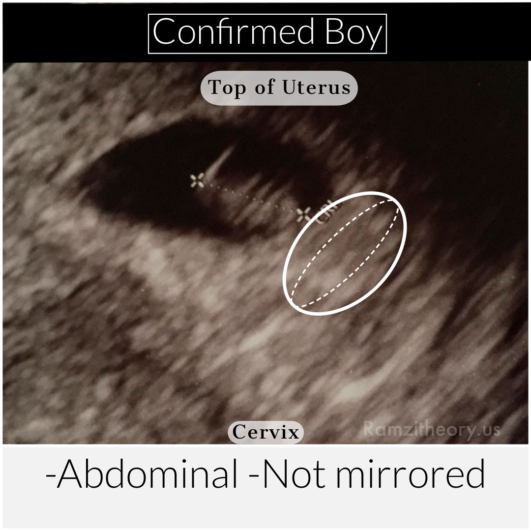
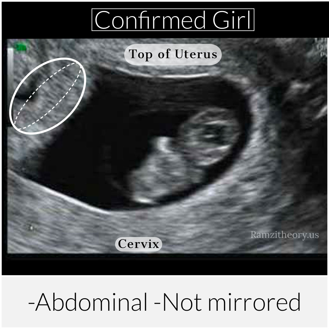
The best way to understand some of the ways the Ramzi theory ultrasounds should be read is to use some visual examples. We can see how each image differs from shape, position and the annotations or lack there of which give us the information necessary to make educated decisions. In the following Ramzi theory examples we will see different types of ultrasound, all marked abdominal or transvaginal and we can compare the confirmed Ramzi theory results.

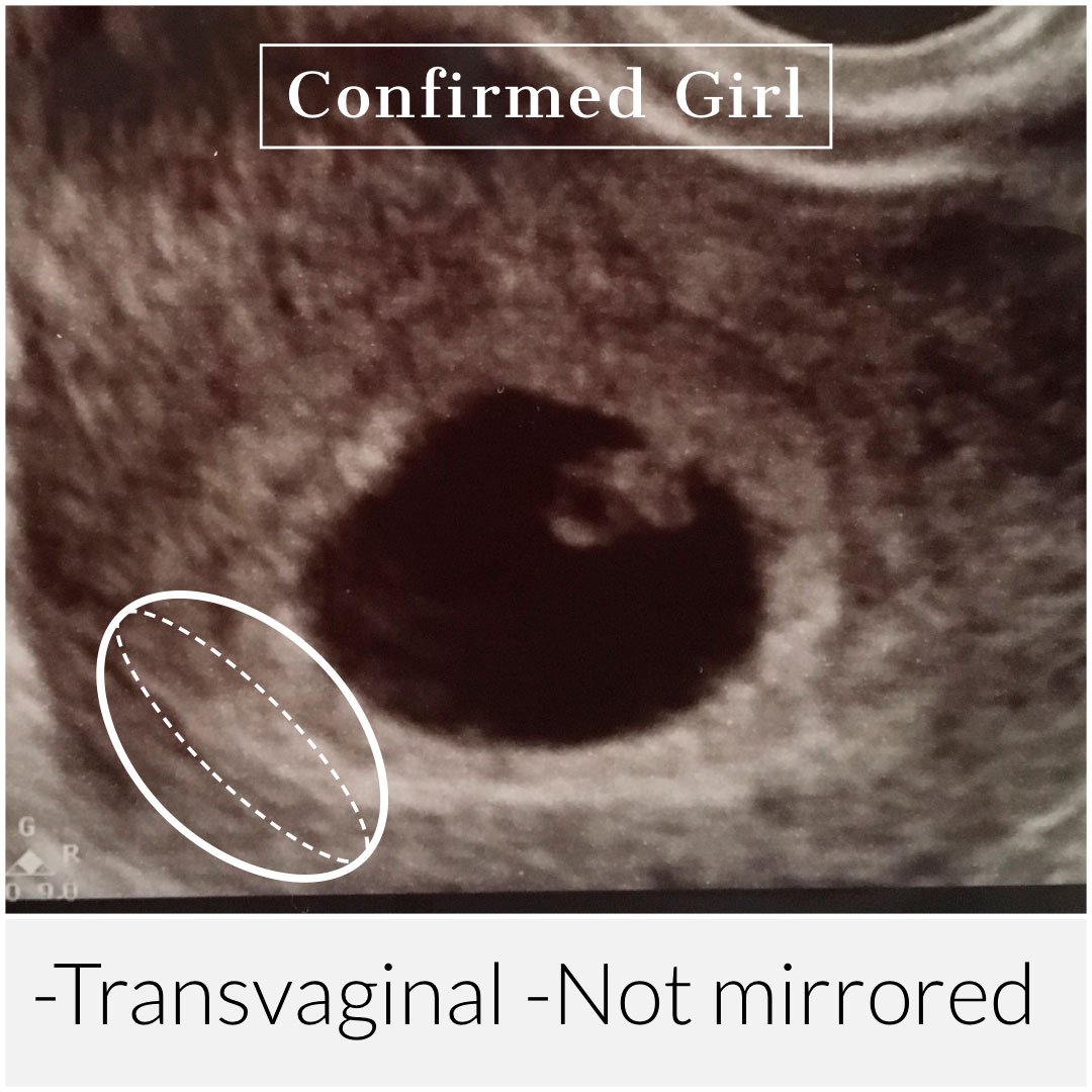

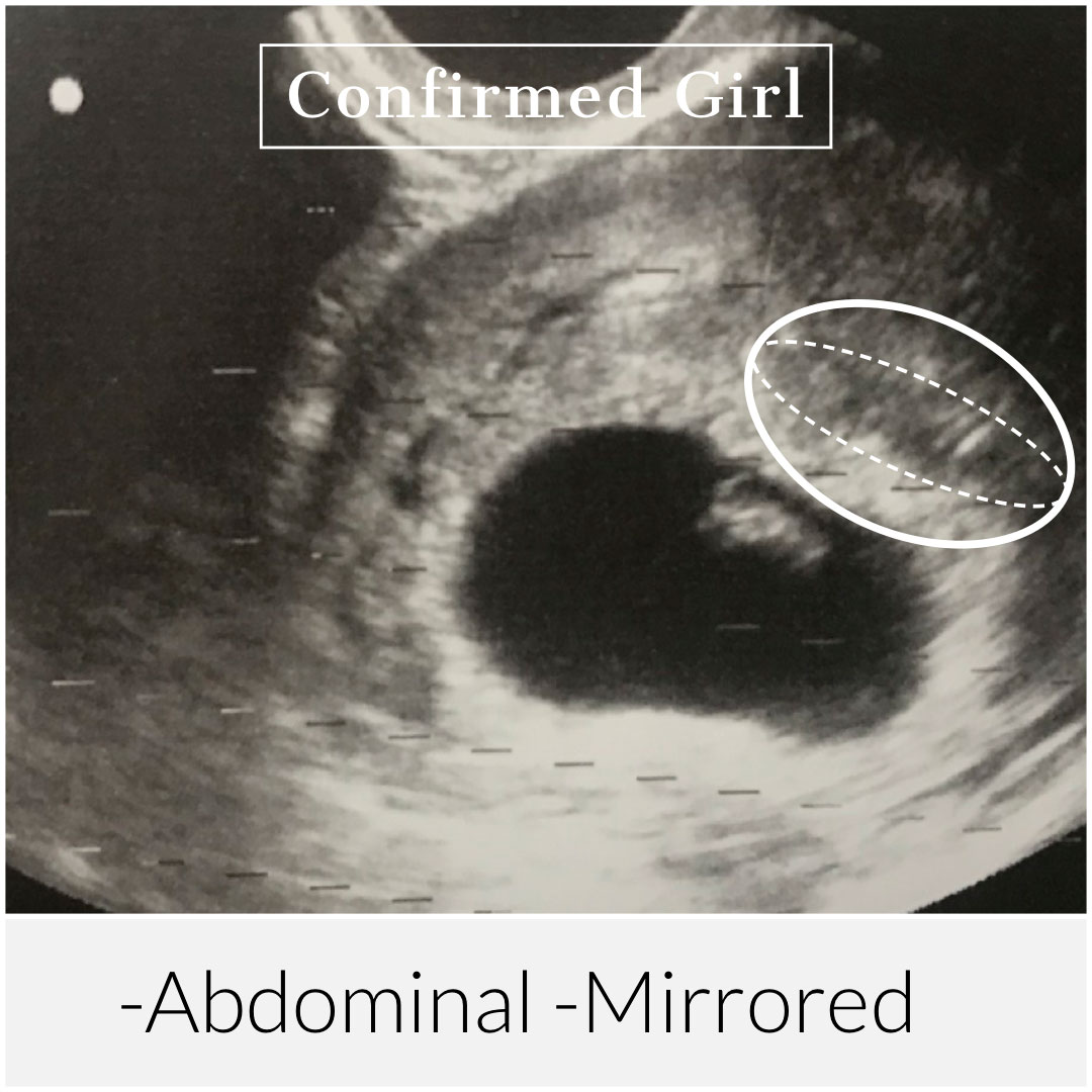
In the above examples we can see how Ramzi theory transvaginal ultrasound examples show the possibilities between mirrored and true-to-side readings depending on the annotations or word confirmation given by the ultrasound technician. By not applying the same flipped or not flipped rule that is talked about in most non-professional articles or websites, our professional team is able to provide a correct prediction with a higher overall accuracy rate.
The more Ramzi theory pictures our clients see, the more they notice our specialized techniques and how they increase our accuracy.
There are other markers that are important to noticed when analyzing Ramzi ultrasound pictures. Keeping the top of the uterus differentiated from the start of the cervix is key. Finding the direction of blood flow can provide highly helpful insight. Ultrasound technician computer annotations can make all the difference when in doubt of whether an image should be flipped or stay true-to-side. Below we provide a variety of Ramzi theory ultrasound examples that show different views and essential markings that are not to be missed.
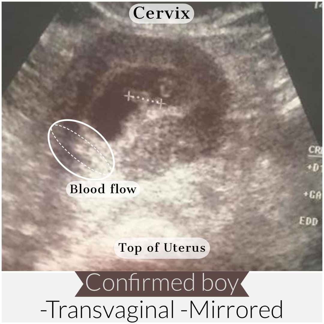
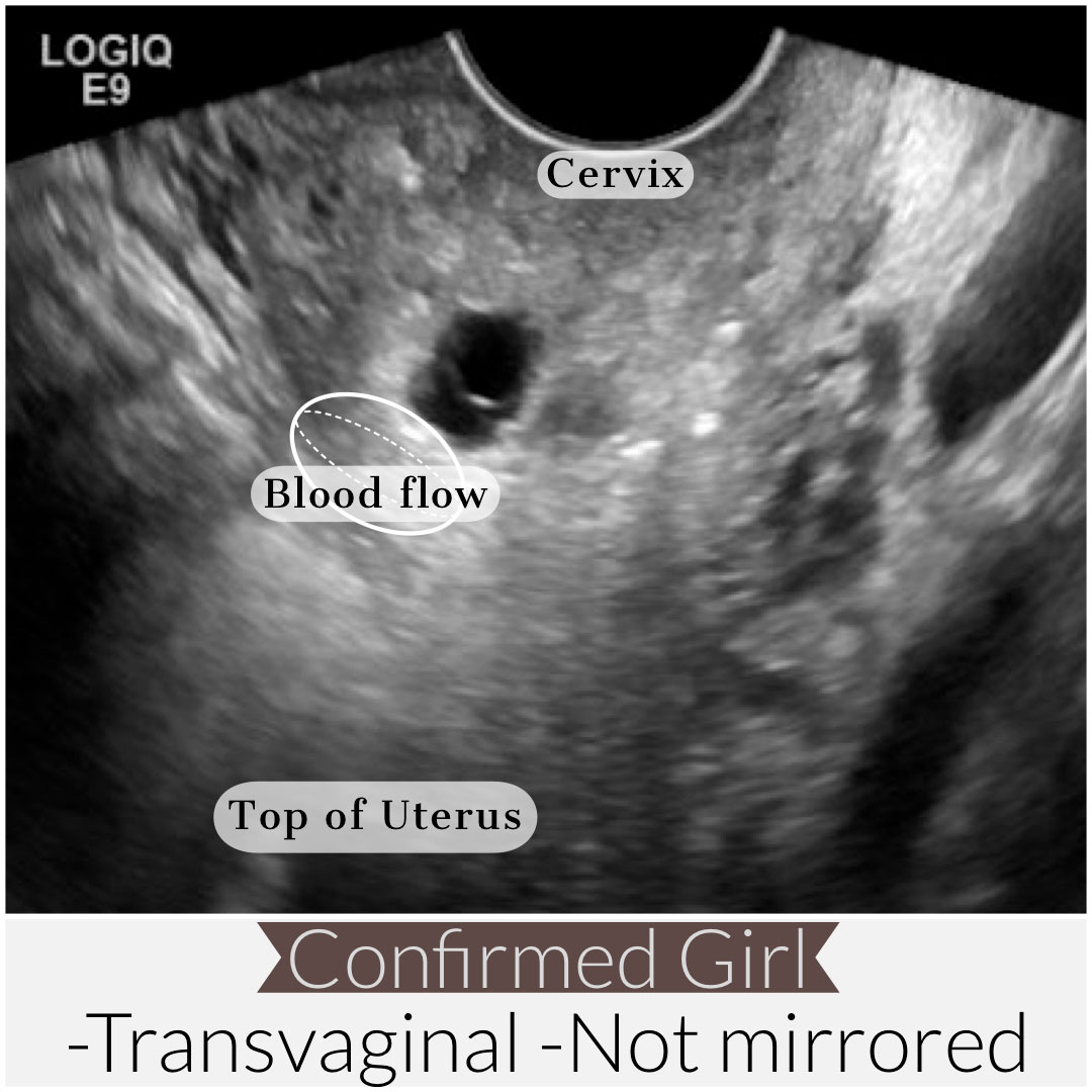
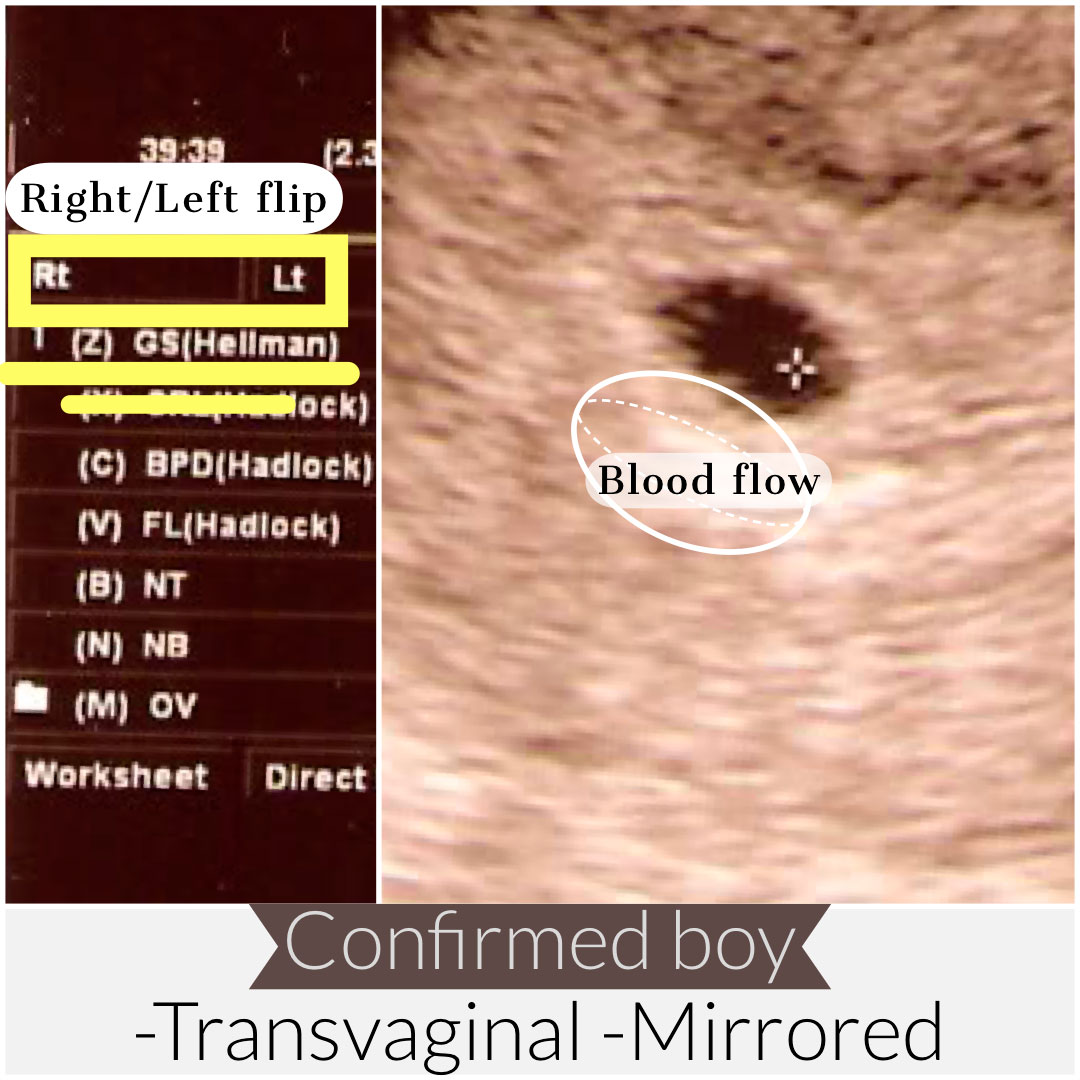
Studying the above Ramzi theory transvaginal ultrasound examples we can be sure that the popular thought that all transvaginal ultrasound are true to side is not correct. We can observe several Ramzi theory ultrasound examples that prove that believe to be incorrect. In the third of the Ramzi theory examples, we can see a confirmed boy ultrasound from a mirrored transverse ultrasound picture. This Ramzi theory example can be easily analyzed by looking at the annotations provided at the top left corner of the ultrasound picture. The markings “Rt-Lt” show the flipped sides of the image and thus corroborates our Ramzi theory prediction.
In other Ramzi theory ultrasound examples we can see other annotations such as the abbreviations “TSV” or “Transv” to indicate transverse scanning plane ultrasounds. Also common is “SG” or Sagt” for a sagittal scanning plane. Knowing the scanning plane of an ultrasound can highly increase the accuracy of a Ramzi theory prediction. If in doubt, it is always recommended to ask the ultrasound technician to write down as many annotations as possible, or provide verbal information of the techniques used during an ultrasound.
Lastly we can analyze some Ramzi theory abdominal ultrasound examples that show that even abdominal ultrasounds can sometimes not be mirrored. Because flipping the sides of an ultrasound is a feature that the technician can use at anytime, it’s important to know what to look for. The following Ramzi theory abdominal ultrasounds provide a variety of possible examples.
